Observing a single Li-ion-column at atomic resolution using aberration-corrected annular-bright-field microscopy
Date:23-09-2011 Print
Transmission electron microscopes (TEM) are amongst the most powerful tools for observing the microstructure of materials at high spatial resolution. The trend of modern scientific development has required characterizing the physical and chemical properties of materials and solving specific problems in condense matter physics at atomic resolution (better than 1 angstrom). However, because of the presence of lens aberrations, in particular the spherical aberration, the resolution of a microscope is usually limited to be close to 2 angstrom using conventional approaches. Different from previous accesses to improve the microscope resolution, which relies mainly on intensive post processing and complicated image simulations, the first Cs (spherical aberration coefficient) corrector was demonstrated at the end of last century. Cs-corrected transmission electron microscopy has since then made it possible to acquire structural information of materials directly with sub-angstrom resolution. However, a critical challenge remains in TEM that the light elements (such as hydrogen, lithium etc.) can not be resolved directly by conventional methods due to their poor electron scattering cross sections.
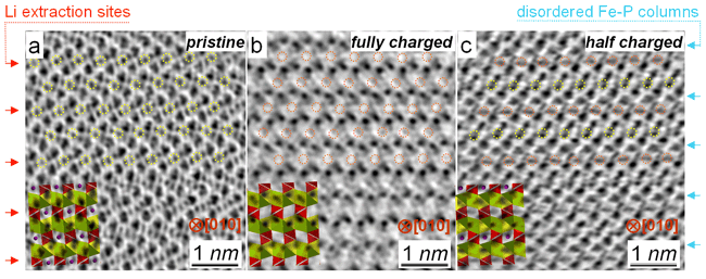
Recently, Prof. Lin Gu, Prof. Xiaofeng Duan and Dr. Xiaoqing He at the Beijing National Laboratory for Condensed Matter Physics, The Institute of Physics, Chinese Academy of Sciences (IOP, CAS) in close collaboration with Prof. Hong Li, Prof. Yong-Sheng Hu and Prof. Liquan Chen (IOP, CAS), together with scientists from the Max Plank Institute for Solid State Research, Japan Fine Ceramic Center and The University of Tokyo, have developed an annular-bright-field scanning transmission electron microscopy, which can image lithium atoms directly in cathode materials e.g. LiFePO4 (http://pubs.acs.org/doi/abs/10.1021/ja109412x). Corresponding relationship between experimental parameters and image contrast was also discussed systematically (Mater. Express 1, 43-50, 2011). In addition, it is found that in partially delithiated LiFePO4 the remaining lithium ions preferentially occupied every second layer, analogous to the staging phenomenon in graphene. This result challenges previous core-shell or phase boundary model, which has strong implications to unravel the underlying mechanism of lithium storage and understand the “staging phenomenon”. These results were supported subsequently by theoretical calculations by Malik Rahul et al. (Nature Mater., 10, 587-590, 2011).
This work was supported by Chinese Academy of Sciences and “863” project (2009AA033101) from The Ministry of Science and Technology.
Recently, Prof. Lin Gu, Prof. Xiaofeng Duan and Dr. Xiaoqing He at the Beijing National Laboratory for Condensed Matter Physics, The Institute of Physics, Chinese Academy of Sciences (IOP, CAS) in close collaboration with Prof. Hong Li, Prof. Yong-Sheng Hu and Prof. Liquan Chen (IOP, CAS), together with scientists from the Max Plank Institute for Solid State Research, Japan Fine Ceramic Center and The University of Tokyo, have developed an annular-bright-field scanning transmission electron microscopy, which can image lithium atoms directly in cathode materials e.g. LiFePO4 (http://pubs.acs.org/doi/abs/10.1021/ja109412x). Corresponding relationship between experimental parameters and image contrast was also discussed systematically (Mater. Express 1, 43-50, 2011). In addition, it is found that in partially delithiated LiFePO4 the remaining lithium ions preferentially occupied every second layer, analogous to the staging phenomenon in graphene. This result challenges previous core-shell or phase boundary model, which has strong implications to unravel the underlying mechanism of lithium storage and understand the “staging phenomenon”. These results were supported subsequently by theoretical calculations by Malik Rahul et al. (Nature Mater., 10, 587-590, 2011).
 |
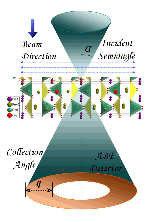
| Fig. 1(a) Experimental set-up of the annular-bright-field (ABF) imaging condition. An incident sub-angstrom probe with convergent semiangle α is scanning on the sample. ABF micrographs can be then obtained via changing the geometry of an annular detector to collect electrons at an angle ranging q within the bright field disc. (Image by Lin Gu et al.) |
 |
| Fig. 2(a) An ABF micrograph along [010] direction for raw LiFePO4, where black dots denote locations of atoms. The lithium ions (yellow circles) are clearly visible. (b) For fully charged state, the lithium ions are extracted completely, and the delithiated sites are marked by red circles. (c) For partially charged sample, the remaining lithium ions (yellow circles) preferentially occupied every second layer, presenting a “staging phenomenon”. (Image by Lin Gu et al.) |


